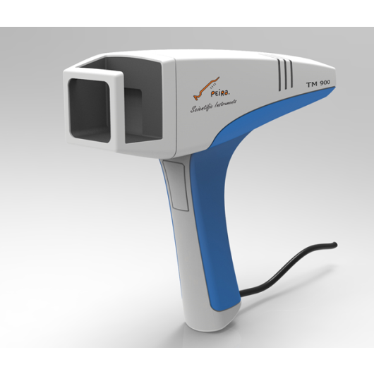
Tumor scanner - demo product
The first version of the Peira scanner is a post-demonstration model. The device was serviced and calibrated by the manufacturer. We offer the possibility of purchasing it at a reduced price, after a week of free trial by the customer.
TM900 is a complete platform; besides the handled device, the TM900 comes with a touch screen panel PC with the measurement software.
The measurement software allows visualizing the tumor topography and the analyzed surface.
Important tumor features such as height, width, depth and volume are automatically calculated. Previous measurements of the animals are shown on the screen to allow instantaneous folllow-up of tumore volume over tie. The software interface also allows coupling with other hardware such as balances, RFID scanners, ... to allow for complementary measurements. An export function is foreseen to export the data to the management software for further ananlysis.
The management software allows for a complete data management of your data; you can defne experiments, assign animals to groups (randomization), visualize and reanalyze the tumors and make plots of the data. Export to e.g. exel is foreseen.
Whay the TM900?
-
Visualization in 3D of the tumor topography. This allows for visual inspection of the tumor shape and morphology over time and flagging of the necrotic or inflamed tumors.
-
Data integrity and traceability. The software allow for storage, ananlysis, visualization and management of the data, which is essential for robust data follow-up and QA-requirements
-
Validation. High resolution 3D images are acquired, allowing precise volume estimations and validation of the measured surface. Especially oddly shaped or thin tumors benefit from TM900 volume estimations.
Watch more
- Measurement range (X - Y): 25 mm - 25 mm
- Maximum tumor size (L x W x H): 20 mm x 20 mm x 20 mm
- Accuracy per measured 3D point: < 0,3 mm
- Stereo capture time: ~ 0,1 s
- Device, panel-pc interface
- USB 2.0
- Camera 1600 x 1200 pixel (2 MP)
- Projector 300 x 300 pixels, 532 nm (green for optimal contrast)
- Camera/projector working distance: 50 mm
Mendel University in Brno, Czech Republic
International Institute of Molecular and Cell Biology, Warsaw, Poland
Publications by Memorial Sloan Kettering Cancer Center
- Aberrant Expression of ERG Promotes Resistance to Combined PI3K and AR Pathway Inhibition through Maintenance of AR Target Genes
- ImmunoPET Predicts Response to Met-targeted Radioligand Therapy in Models of Pancreatic Cancer Resistant to Met Kinase Inhibitors
- Bispecific antibody does not induce T-cell death mediated by chimeric antigen receptor against disialoganglioside GD2
- A PET Imaging Strategy for Interrogating Target Engagement and Oncogene Status in Pancreatic Cancer
- GREB1 amplifies androgen receptor output in human prostate cancer and contributes to antiandrogen resistance
- Regulation of the glucocorticoid receptor via a BET-dependent enhancer drives antiandrogen resistance in prostate cancer
- SOX2 promotes lineage plasticity and antiandrogen resistance in TP53– and RB1-deficient prostate cancer
- Abstract B38: Tetravalent bispecific antibodies specific for HER2 and disialoganglioside GD2 to engage polyclonal T cells for osteosarcoma therapy
- Pretargeted Radioimmunotherapy Based on the Inverse Electron Demand Diels-Alder Reaction.
- A rectal cancer model establishes a platform to study individual responses to chemoradiation
- A potent tetravalent T-cell–engaging bispecific antibody against CD33 in acute myeloid leukemia
- Tumor-Specific Zr-89 Immuno-PET Imaging in a Human Bladder Cancer Model
- Development of a tetravalent anti-GPA33/anti-CD3 bispecific antibody for colorectal cancers
- FOXA1 mutations alter pioneering activity, differentiation, and prostate cancer phenotypes
- Interdomain spacing and spatial configuration drive the potency of IgG-[L]-scFv T cell bispecific antibodies
Publications by Hainan Medical College
- 3′-epi-12β-hydroxyfroside, a new cardenolide, induces cytoprotective autophagy via blocking the Hsp90/Akt/mTOR axis in lung cancer cell
- Coroglaucigenin induces senescence and autophagy in colorectal cancer cells
- Genetically engineered drug rhCNB induces apoptosis and cell cycle arrest in both gastric cancer cells and hepatoma cells
- Induction of enhanced immunogenic cell death through ultrasound-controlled release of doxorubicin by liposome-microbubble complexes
- Toxicarioside O Inhibits Cell Proliferation and Epithelial-Mesenchymal Transition by Downregulation of Trop2 in Lung Cancer Cells
Institute of Biomedical Engineering, University of Oxford
Taichung Veterans General Hospital, Taichung, Taiwan
• City University of Hong Kong
• Hunter College, CUNY*
• Vanderbilt University Medical Center*
• Hainan Medical University*
• Howard Hughes Medical Institute*
• Memorial Sloan Kettering Cancer Center*
• Cancer Center Amsterdam
• GlaxoSmithKline
• Imec
• K.U.Leuven
• Neuro-Electronics Research Flanders
• Pepric
• PharmaVize
• reMYND
• University Ghent
• SEPS Pharma
• Vlaams Instituut voor Biotechnologie
• University Antwerp
• ThromboGenics
• Janssen Pharmaceutica

